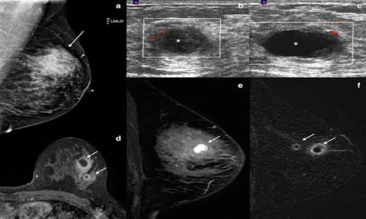
A callback after a routine mammogram can transform an ordinary week into days filled with uncertainty. While most breast abnormalities detected through imaging turn out to be benign, the period between discovery and diagnosis requires careful navigation of medical procedures, emotional responses, and practical decisions about next steps.
What Imaging Actually Detects
Mammograms, ultrasounds, and MRI scans identify areas that differ from surrounding breast tissue. These differences might appear as dense clusters of microcalcifications, irregular masses, or changes in tissue architecture. Radiologists classify findings using the BI-RADS system, which assigns a score from 0 to 6 based on the level of concern. A BI-RADS 3 indicates probably benign findings with short-interval follow-up recommended, while BI-RADS 4 and 5 suggest tissue sampling is necessary to rule out malignancy.
Microcalcifications represent tiny calcium deposits that form in breast tissue. While most are harmless, certain patterns-particularly fine, irregular clusters-warrant closer examination. Masses present different challenges depending on their borders, shape, and density. A well-circumscribed, round mass with smooth edges typically suggests a benign cyst or fibroadenoma, while spiculated or irregular borders raise greater concern.
The Diagnostic Pathway After Abnormal Results
When initial imaging reveals an abnormality, diagnostic mammography provides additional views with magnification and compression techniques that better characterize the finding. Targeted ultrasound helps distinguish fluid-filled cysts from solid masses and provides real-time visualization of tissue structure. These additional imaging studies sometimes resolve the question entirely-a clearly benign simple cyst requires no further action beyond routine monitoring.
However, when imaging alone cannot establish whether an abnormality is benign or malignant, tissue sampling becomes necessary. The type of biopsy recommended depends on what imaging detected, where it sits within the breast, and what visualization method provides the clearest guidance for accessing it. Understanding these factors helps patients prepare mentally and practically for the procedure ahead.
Precision-Guided Tissue Sampling Methods
When mammography identifies suspicious microcalcifications or masses visible only on mammogram views, a specialized approach ensures accurate sampling. A stereotactic biopsy breast procedure uses three-dimensional mammographic imaging to pinpoint the exact location of abnormal tissue. The patient lies prone on a specialized table with the breast positioned through an opening, allowing imaging from multiple angles to calculate precise coordinates. This method proves particularly effective for sampling calcifications that cannot be felt or seen with ultrasound, delivering millimeter-level accuracy while the patient remains still throughout the procedure.
Ultrasound-guided biopsy serves as the primary method for palpable masses or those clearly visible on ultrasound imaging. The real-time visualization allows the radiologist to watch the needle enter the tissue and adjust position as needed. For abnormalities detected only on MRI, MRI-guided biopsy provides the necessary visualization, though these procedures take longer and require specialized equipment available at advanced imaging centers.
What Happens During and After Biopsy
Core needle biopsies remove several tissue samples, each about the size of a grain of rice, through a hollow needle. Local anesthesia numbs the area, and patients typically describe pressure or a snapping sensation rather than pain. The entire procedure usually takes 30 to 60 minutes, with most of that time spent on imaging and positioning rather than actual sampling. A small titanium marker clip is often placed at the biopsy site to mark the location for future reference, particularly important if the area would be difficult to relocate on subsequent imaging.
Post-procedure care involves ice application and wearing a supportive bra for several days. Bruising and tenderness are normal and typically resolve within a week. Most patients resume normal activities the next day, avoiding only strenuous exercise or heavy lifting for 24 to 48 hours. Results arrive within three to seven days, during which uncertainty often peaks despite the relatively short wait.
Interpreting Pathology Results
Pathology reports classify findings into several categories. Benign results include normal tissue, fibrocystic changes, fibroadenomas, or benign calcifications. These findings typically require only routine screening going forward. Atypical results indicate cells that show abnormal features but do not meet criteria for malignancy-these often warrant surgical excision because they carry increased cancer risk and sometimes upgrade to cancer upon removing the entire area.
Malignant results identify the specific type of breast cancer, its hormone receptor status, and other characteristics that guide treatment planning. Even within cancer diagnoses, considerable variation exists in prognosis and treatment approach. Ductal carcinoma in situ represents non-invasive cancer confined to milk ducts, while invasive cancers have penetrated surrounding tissue. The pathology report provides the roadmap for the medical team to develop an individualized treatment strategy.
Managing the Emotional Weight
The period between detecting an abnormality and receiving definitive results creates psychological strain that rivals physical discomfort. Anxiety, sleep disruption, and intrusive thoughts about worst-case scenarios are normal responses to medical uncertainty. Many patients find that structured coping strategies help: scheduling the biopsy quickly rather than prolonging the wait, arranging practical support for procedure day, and identifying one trusted person to process results with rather than crowd-sourcing opinions.
Healthcare navigation shares common elements across medical specialties-whether selecting providers for routine care or coordinating complex diagnostic workups, patients benefit from understanding typical processes and asking informed questions. Research shows that hospital-based specialty practices often provide coordinated teams where radiologists, pathologists, and surgeons communicate directly, streamlining the transition from diagnosis to treatment when needed.
Moving Forward After Diagnosis
Benign results bring relief, but patients should maintain regular screening schedules and report new changes promptly. A benign finding today does not prevent future abnormalities, and establishing patterns through consistent imaging helps radiologists detect subtle changes over time. Some benign conditions, particularly atypical hyperplasia, may warrant more frequent monitoring or preventive medications to reduce future cancer risk.
For those facing cancer diagnoses, the initial shock gives way to treatment planning meetings, additional staging tests, and decisions about surgery, radiation, and systemic therapies. Multi-specialty cancer centers coordinate these complex pathways, but patients remain central decision-makers throughout. Asking about expected timelines, treatment sequence, and the rationale behind recommendations helps patients feel more grounded during a disorienting time.
The discovery of a breast abnormality initiates a process rather than delivering immediate answers. Understanding what different imaging modalities reveal, how biopsy techniques match specific findings, and what pathology results mean equips patients to participate actively in their care. While medical uncertainty creates stress, the diagnostic tools available today provide remarkably precise information that guides appropriate treatment while avoiding overtreatment of benign conditions.